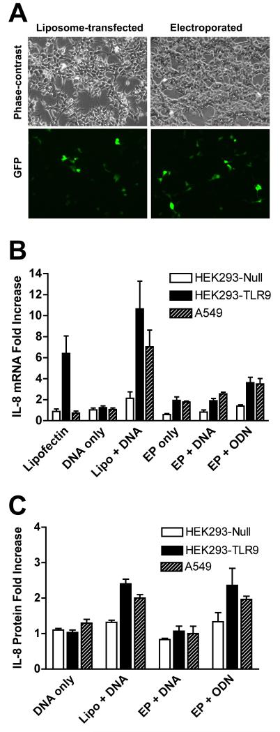
Figure 1. Activation of IL-8 expression in cells with different levels of TLR9.
(A) HEK293 cells were transfected with 5 μg pEGFP-N1 (Clontech) by electroporation or Lipofectin (Invitrogen, Carlsbad, CA). For electroporations, 1 × 10⁶ cells were suspended in DMEM containing 10% fetal bovine serum, placed in cuvette with a 4 mm gap, and electroporated at 160V with a capacitance of 950 μF using a Bio-Rad Gene Pulser II. For liposomal gene delivery, DNA was complexed with 10 μl of Lipofectin, incubated for 30 minutes, diluted into DMEM without serum, and added to cells, as described by the manufacturer (Invitrogen). Transfection efficiency was evaluated 24 hours after transfection. (B) HEK293-TLR9-Null, HEK293-TLR9+ cells (InvivoGen, San Diego, CA) and A549 cells (ATCC) were maintained in DMEM supplemented with 10% fetal bovine serum, kanamyacin, antibiotic/antimyotic solution (Invitrogen, Calsbad CA), and 10 μg/ml blasticidin (HEK293 cells only). Cells were transfected with 5 μg pEGFP-N1 using different conditions as indicated. Alternatively, cells were electroporated in the absence of plasmid DNA, and 10 minutes later, the CpG-containing oligonucleotide ODN M362 (InvivoGen) was added to the cells at a final concentration of 10 μg/ml (EP + ODN). Twenty-four hours later, total RNA was isolated and 500 ng was used for reverse transcription. One-tenth of the cDNA was subjected to quantitative PCR. The expression of IL-8 is normalized to the expression of GAPDH. Fold of increase is over the untreated cells. Mean levels ± SEM are shown (n=4). (C) Twenty-four hours after transfection, IL-8 protein levels in cleared supernatant from 1 ml of culture medium were measured using a human IL-8 ELISA kit (BD Biosciences, San Diego, CA). Mean levels ± SEM are shown (n=6).

