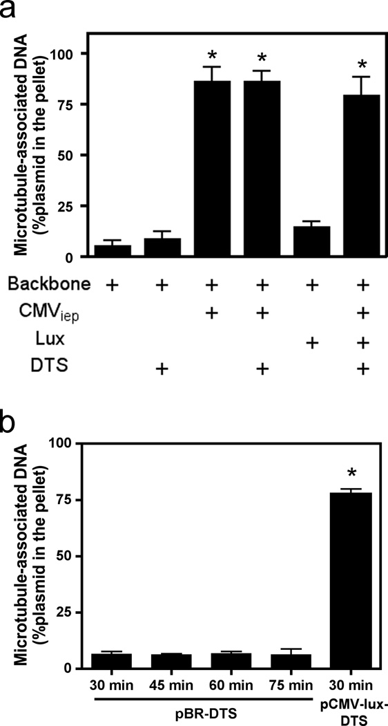
Figure 1. Microtubule spin-down assays showing in vitro interaction of DNA with microtubules.
(a) Quantitative analysis of plasmid elements that associate with microtubules. Plasmids containing different sequence elements (the CMV promoter, the luciferase gene, and/or the DTS) were incubated for 30 minutes with cell extract and taxol-stabilized microtubules and subsequently separated over a glycerol cushion by centrifugation. The plasmid contents of the pellets and supernatants were determined by quantitative PCR, and percentage of DNA in the pellet was determined by comparing DNA content in pelleted fractions versus total DNA in both supernatant and pellet fractions combined. (b) Increased incubation times do not change the ability of the DTS to mediate microtubule interactions. pBR322-DTS was incubated with microtubules in the presence of cell extract for 30, 45, 60, and 75 minutes and subsequently centrifuged and quantified by quantitative PCR as in a. Mean DNA concentrations from three independent experiments, preformed in duplicate, are shown ± st. dev. CMV, Cytomegalovirus; Lux, luciferase gene; DTS, Simian Virus 40 DNA nuclear targeting sequence.

