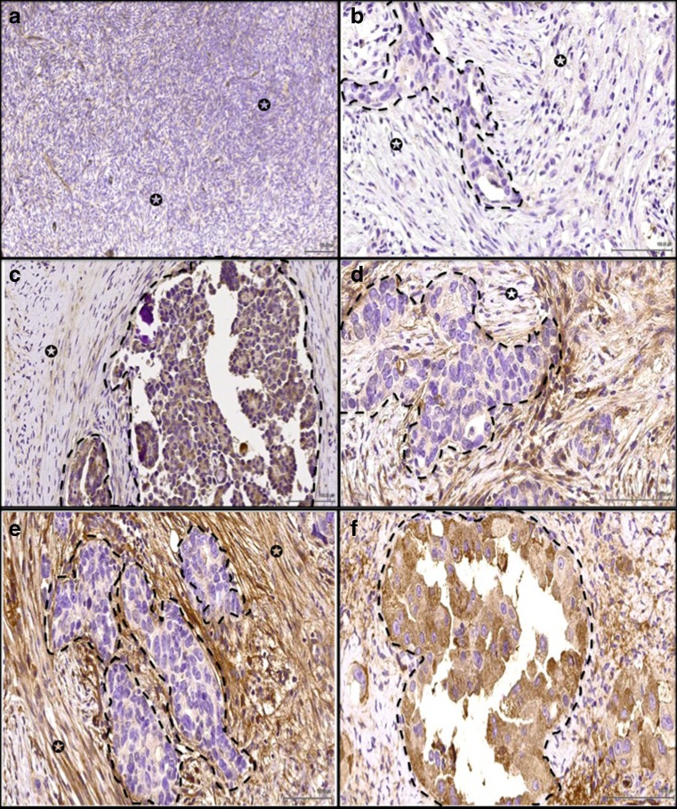
Fig. 1.
a Normal ovary stained with FAP showed stromal cells negative for FAP (arrow indicate stroma). Example of ovarian cancer stained for FAP antibody where (arrow indicates stromal cells, dotted lines highlight the cancer cells). In (b), the stromal cells showed negative expression; c Weak expression. d Moderate expression and e Strong expression. Ovarian cancer cells were strong positive (f). ×40

