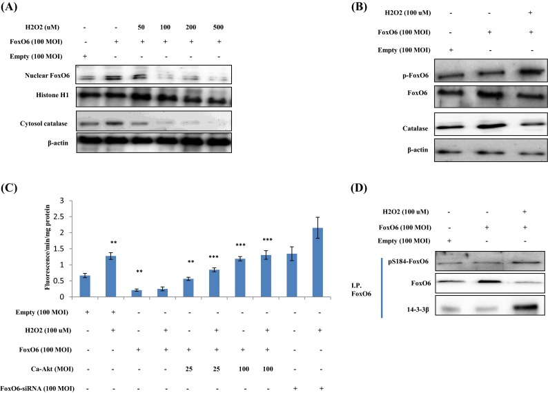
Fig. 6.

Enhancement of FoxO phosphorylation in H2O2-treated HEK293T cells. HEK293T cells were treated with H2O2 at various concentrations, and FoxO6 levels were determined by Western blotting. Samples loaded on sample gels were probed with β-actin and histone H1. a Levels of nuclear FoxO6 and cytoplasmic catalase were noticeably diminished after treatment with H2O2 at 50 to 500 μM. b Nuclear phosphorylated FoxO6 and catalase levels were noticeably decreased by 100 μM H2O2 in FoxO6-transfected (100 MOI) cells. c Quantitative analysis was performed by measuring DCFDA fluorescence after treating cells with vehicle or 100 μM H2O2 in the absence or presence of FoxO6 (100 MOI), CA-Akt (25, 100 MOI), or FoxO6-siRNA (100 MOI) for 1 day. Results of one-factor ANOVA: ** p < 0.01 vs. H2O2 untreated cells; *** p < 0.001 vs. H2O2-treated cells. d Immunoprecipitated FoxO6 was found to be physically associated with 14-3-3β by Western blotting
