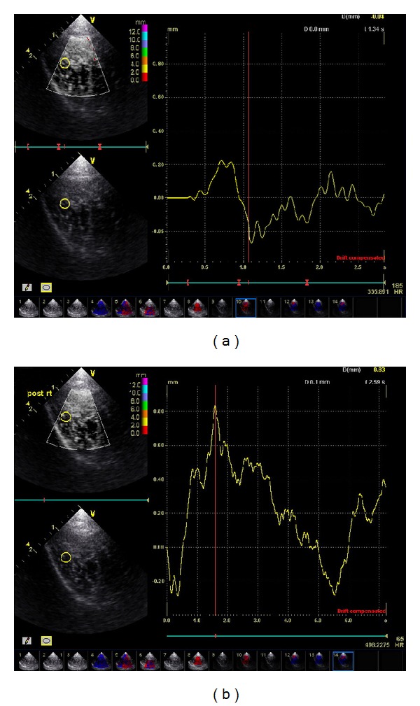
Figure 3.

Tissue displacement on acupoints following needle stimulation before and after de qi. In vivo ultrasonic imaging using a System FiVe (Vingmed) at 7.5 MHz was performed on the healthy subjects at different stages of acupuncture needle stimulation including before de qi and during de qi. Displacements were estimated using the ultrasonic radio-frequency (RF) data. Seventy RF scans were acquired continuously during each experiment at the rate of 13.2 frames per second.
