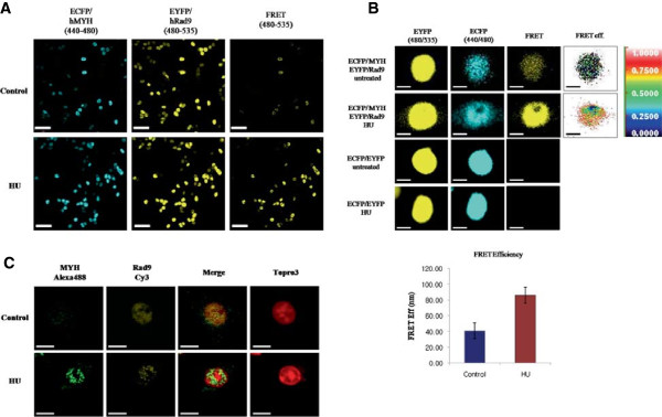
Figure 5.

DNA damage (HU) promotes the interaction between hRad9 and hMYH. (A) Fluorescence resonance energy transfer (FRET) was used to analyze the interaction between over-expressed hRad9 and hMYH after DNA damage. Cells transfected with ECFP and EYFP or ECFP/hMYH and EYFP/hRad9 were treated with or without HU. ECFP, EYFP, and FRET fluorescence was observed with a fluorescence microscope at 440/480 nm (ECFP), 480/535 nm (EYFP), and 440/535 nm (FRET). Scale bar, 50 μm. (B) FRET efficiency was used to quantify the interaction between hMYH and hRad9 after DNA damage. HEK293 cells were transfected with ECFP and EYFP or ECFP/hRad9 and EYFP/hMYH as indicated to the left of the figure. Cells were treated with or without HU. ECFP, EYFP, and FRET fluorescence were measured at the indicated wavelengths. FRET efficiency was quantified in five control cells and five HU-treated cells. Standard error is shown. Scale bar, 10 μm. (C) Immunofluorescence of endogenous hMYH and hRad9. Cells were treated with or without 20 mM HU for 1 h and allowed to recover for 2 h. Cells were stained with antibodies against hMYH (FPG 456, green), hRad9 (FPR 552, yellow), and To-pro®-3 (nuclei, red). Scale bar, 10 μm.
