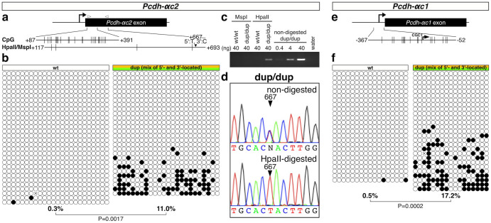
Figure 5. Tandem duplication increases the DNA methylation of Pcdh-αc2 and Pcdh-αc1.
(a) Schematic representation of Pcdh-αc2; the positions of CpGs and HpaII/MspI sites are shown to scale by vertical lines. (b) Results of bisulfite sequencing. Each circle represents a methylated (black) or unmethylated (white) CpG dinucleotide. Each row represents a single clone. A primer set was designed to amplify the region corresponding to the 5′ region of the exon, which has the same sequence in the 5′- and 3′-located duplicated Pcdh-αc2 isoforms. The percentage below each methylation pattern indicates the CpG methylation rate for the region. C, D, Discrimination of the DNA methylation between the 5′- and 3′-located duplicated Pcdh-αc2 isoforms by HpaII digestion-mediated analysis. (c) Electrophoresis results. D, Example chromatograms showing that the HpaII-resistant fraction contained the region of the 5′-located duplicated Pcdh-αc2 exon. E, Schematic representation of Pcdh-αc1; positions of CpGs are shown to scale by vertical lines. F, Results of bisulfite sequencing. A primer set was designed to amplify the region corresponding to the Pcdh-αc1 promoter, which has the same sequence in the 5′- and 3′-located Pcdh-αc1 duplicates. The percentage below each methylation pattern indicates the CpG methylation rate for the region.

