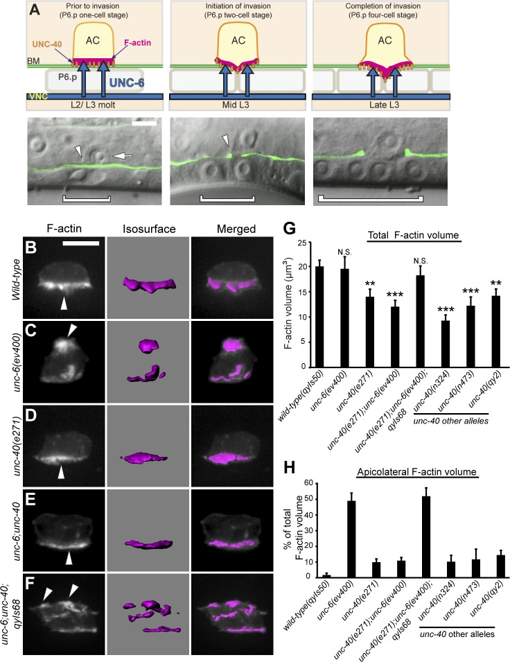
Figure 1.
UNC-40 is mispolarized and active in the absence of UNC-6. Anterior is left; ventral is down. (A) AC invasion in C. elegans (top, schematic; green, differential interference contrast [DIC] microscopy with basement membrane marker laminin::GFP; bottom, overlay). During the L2/L3 molt (left), the AC (bottom, arrow) is attached to the basement membrane (BM; arrowhead) over the P6.p vulval precursor cell (bracket outlines nucleus, bottom). UNC-6 (netrin; top, blue arrows) secreted from the ventral nerve cord (VNC) polarizes UNC-40 (DCC; orange ovals) and F-actin (magenta) to the AC’s basal, invasive cell membrane. After P6.p divides (middle, P6.p two-cell stage), a protrusion breaches (bottom, arrowhead) and then removes basement membrane, and moves between the central P6.p granddaughter cells (right, P6.p four-cell stage). (B–F) Fluorescence (left), corresponding dense F-actin network (isosurface, middle), and overlay (right). (B) In wild-type animals, F-actin (visualized with cdh-3 > mCherry::moeABD) was polarized to the basal membrane (arrowhead). (C) In unc-6 mutants, F-actin was mislocalized to the AC’s apical and lateral membranes (arrowhead). (D) In unc-40 mutants, F-actin volume was reduced but polarized (arrowhead). (E) In unc-6; unc-40 double mutants, F-actin was reduced but polarized (arrowhead), comparable to unc-40. (F) In unc-6; unc-40; qyIs68[cdh-3 > unc-40::GFP] animals, F-actin was mislocalized (arrowheads), resembling unc-6. (G and H) The total volume of F-actin and the percentage that localized apicolaterally, respectively, at the P6.p four-cell stage (n ≥ 15 per genotype). *, P < 0.05; **, P < 0.01; ***, P < 0.001. N.S., no significant difference (P > 0.05, Student’s t test). Error bars indicate ± SEM. Significant differences relative to wild-type are indicated. Bars, 5 µm.

