Figure 3.

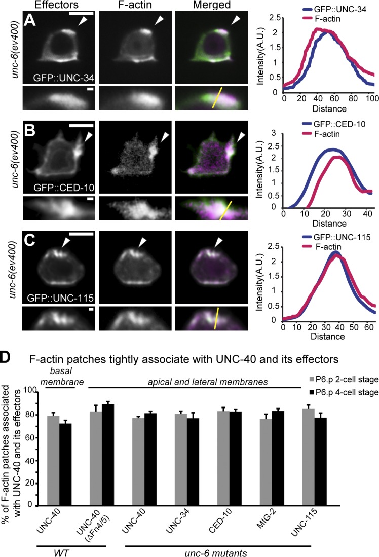
F-actin colocalizes with UNC-40 effectors in the absence of UNC-6. Anterior is left; ventral is down. (A–C) All animals were examined at the P6.p two-cell stage. UNC-40 effectors (left), F-actin (middle), overlay (right), and magnifications (below) are shown. Colocalization graphs (far right) show areas of colocalization (arrowheads; measured along yellow lines in insets; fluorescent intensity is plotted in arbitrary units and distances are given in pixels). In unc-6 mutants, the UNC-40 effectors, GFP::UNC-34 (Ena/VASP), GFP::CED-10 (Rac), and GFP::UNC-115 (abLIM; green), were all colocalized (arrowheads) with F-actin (magenta) at the AC’s apical and lateral membranes. (D) The percentage of the F-actin patches that were associated with the patches of UNC-40::GFP, UNC-40(ΔFN4/5)::GFP, GFP::UNC-34, GFP::CED-10, GFP::MIG-2 (Rac), and GFP::UNC-115 (n ≥ 15 for each stage per genotype). Error bars indicate ± SEM. No significant differences (P > 0.05, Student’s t test) compared with wild-type UNC-40/F-actin were observed. Bars: (main panels) 5 µm; (magnified insets) 0.5 µm.
