Figure 10.
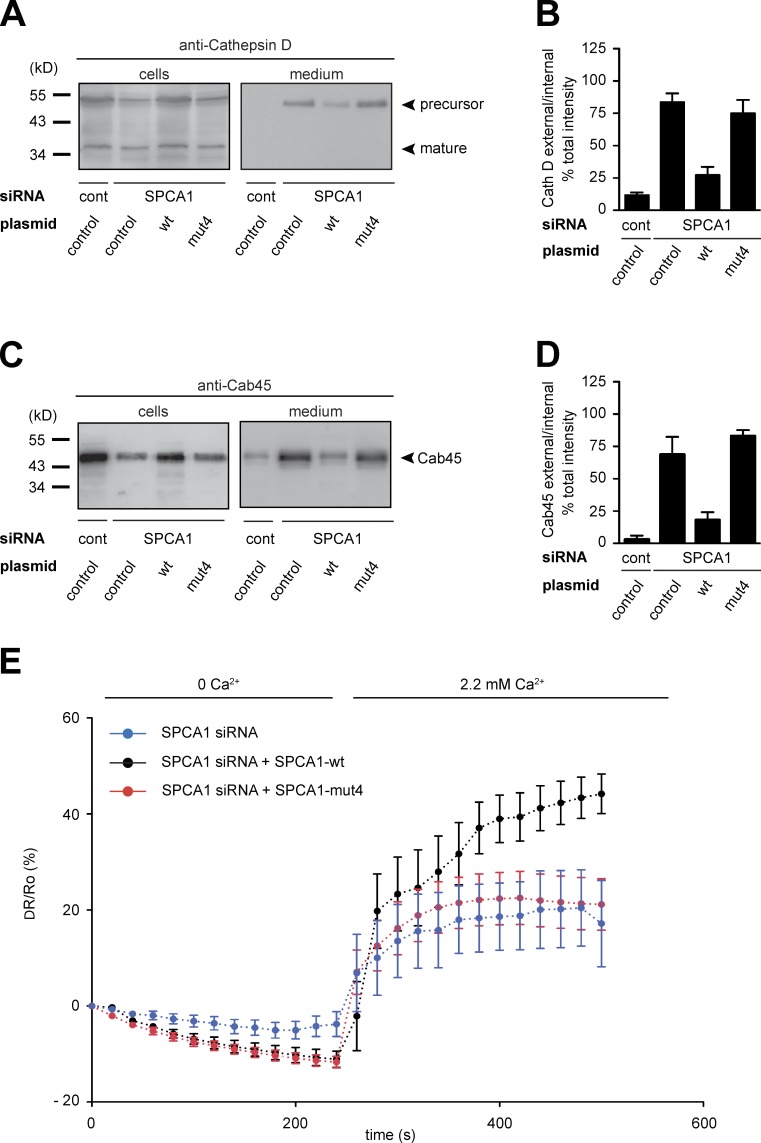
A SPCA1 point mutant impairs TGN Ca2+ uptake and secretory cargo sorting. (A–D) HeLa cells expressing SPCA1-wt and mutants were treated with control and SPCA1 siRNAs. Media and lysates from these cells were Western blotted with Cathepsin D (A) and Cab45 (C) antibodies. Western blots from three independent experiments were quantified by densitometry. Bar graphs represent the densitometry values of external Cathepsin D (B) and Cab45 (D) normalized to internal Cathepsin D and Cab45 values, respectively. (E) HeLa cells expressing either siRNA-resistant SPCA1-wt-HA or -mut4-HA were transfected with SPCA1 siRNA and subsequently with Go-D1-cpv. Ca2+ entry into the TGN was analyzed in wt (n = 8) or mut4 (n = 15)-expressing cells. Fluorescent signals reflecting TGN [Ca2+] were presented as ΔR/R0, where R0 is the value obtained before addition of 2.2 mM Ca2+ to the cell’s bathing solution.

