Figure 4.
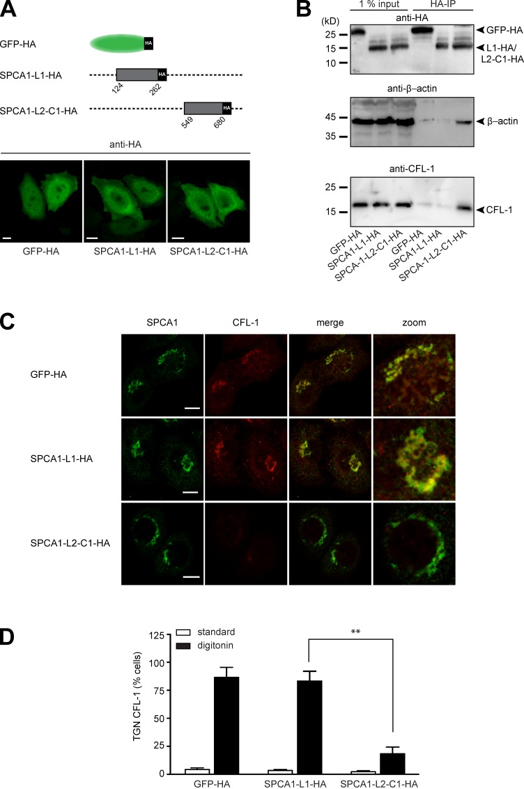
The interaction of CFL-1 and actin with SPCA1-L2-C1 is crucial for protein sorting at the TGN. (A, top) Schematic representation of GFP-HA (aa 1–239), SPCA1-L1-HA (aa 124–252), and SPCA1-L2-C1-HA (aa 549–680) fusion constructs cloned into the retroviral expression vector pLPCX. (A, bottom) HeLa cells stably transfected with GFP-HA, SPCA1-L1-HA, or SPCA1-L2-C1-HA were visualized with an HA antibody and analyzed by confocal microscopy. (B) HeLa cells stably transfected with GFP-HA, SPCA1-L1-HA, or SPCA1-L2-C1-HA were lysed, and lysates were incubated with µMACS anti-HA magnetic microbeads. GFP-HA–, SPCA1-L1-HA–, and SPCA1-L2-C1-HA–associated proteins were eluted, separated by SDS-PAGE, and analyzed by Western blotting using HA, CFL-1, or β-actin antibodies, respectively. (C) HeLa cells stably expressing GFP-HA, SPCA1-L1-HA, and SPCA1-L2-C1-HA were incubated for 2 h at 20°C and subsequently permeablized with digitonin, washed, and then fixed with formaldehyde before incubation with anti-SPCA1 (green) or anti–CFL-1 (red) antibodies. (D) TGN localization of CFL-1 under different conditions in Triton X-100–permeabilized (images in Fig. S2) versus digitonin-permeabilized cells was determined by counting 100 cells per condition in three independent experiments. Bars, 5 µm.

