Figure 6.
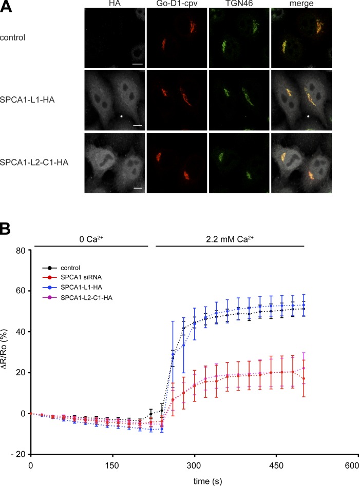
Overexpression of SPC1-L2-C1 inhibits TGN Ca2+ influx. (A) HeLa cells were transfected with the TGN-specific Ca2+ FRET sensor Go-D1-cpv and a control plasmid, SPCA1-HA, or SPCA1-L2-C1-HA. Cells were fixed, stained with anti-HA and anti-TGN46 antibodies, and analyzed by fluorescence microscopy. Bars, 5 µm. (B) HeLa cells were transfected with Go-D1-cpv and a control plasmid, or SPCA1 siRNA and a control plasmid, and either SPCA1-L1-HA or SPCA1-L2-C1-HA, respectively. Ca2+ entry into the TGN was measured in Ca2+-depleted cells transfected with control plasmid (n = 23), SPCA1 siRNA (n = 9), SPCA1-L1-HA, or SPCA1-L2-C1-HA (n = 11). Fluorescent signals reflecting TGN [Ca2+] were presented as ΔR/R0, where R0 is the value obtained before addition of 2.2 mM Ca2+ to the cell’s bathing solution. Data are expressed as the mean ± SEM. Mean maximum values measured after readdition of Ca2+ were statistically different between control/SPCA1-L1-HA and SPCA-L2-C1-HA–transfected cells.

