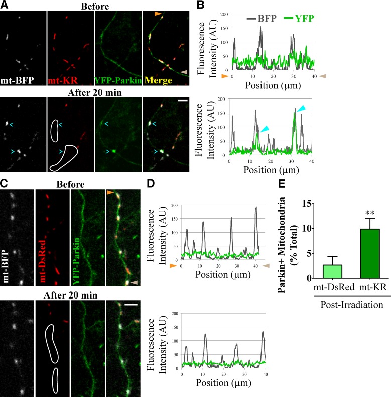
Figure 5.
Parkin is recruited to axonal mitochondria damaged with mt-KR. (A) YFP-Parkin accumulates on a fraction of axonal mitochondria in the outlined area of mt-KR activation. (B) Line scan of the axon in A with cyan arrowheads marking two YFP-Parkin–positive mitochondria. (C and D) YFP-Parkin remained diffuse despite irradiation in the outlined area when mt-DsRed replaced mt-KR. Mitochondria with YFP-Parkin levels more than twice the background were scored as Parkin-positive here and in subsequent figures. Orange and brown arrowheads denote corresponding points in images and line scans. (E) Frequency of YFP-Parkin recruitment to irradiated mitochondria. n = 131–140 mitochondria from 12 transfections. **, P < 0.001. Error bars represent means ± SEM. AU, arbitrary unit. Bars, 5 µm.

