Figure 8.
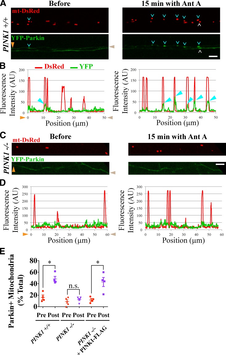
Parkin recruitment to damaged axonal mitochondria requires PINK1. (A–D) YFP-Parkin is recruited to mitochondria of PINK1+/+ (A and B) but not PINK1−/− (C and D) rat hippocampal axons depolarized with 40 µM Antimycin A (Ant A). White and cyan arrowheads denote YFP-Parkin–positive mitochondria; cyan arrowheads point to mitochondria analyzed with line scans in B and D. Orange and brown arrowheads denote corresponding points in images and line scans. (E) Frequency of Parkin recruitment in the indicated genotypes before and 15 min after Antimycin A treatment. n = 87–101 mitochondria from four microfluidic devices per genotype. *, P < 0.05. Error bars represent means ± SEM. AU, arbitrary unit. Bars, 5 µm.

