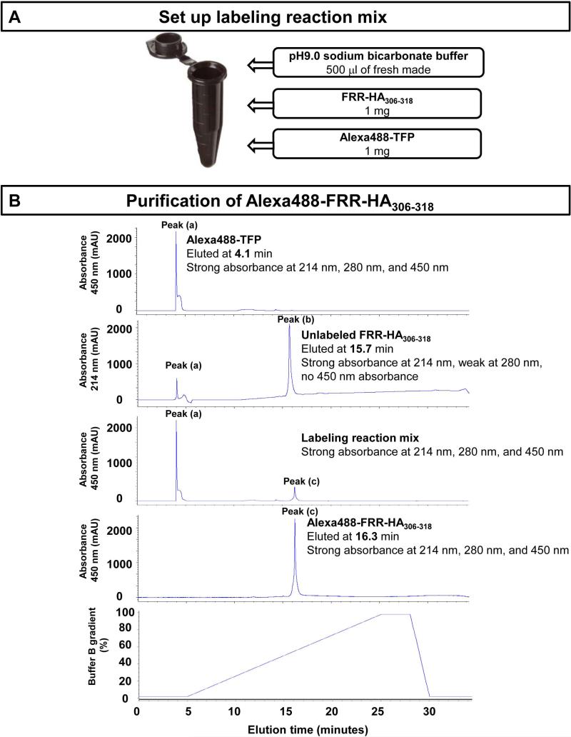
Figure 2. A schematic overview of the steps involved in labeling the probe peptide with a fluorochrome.
(A) Set up the labeling reaction mix and incubate at room temperature for 1 hour. (B) Representative elution profile of fluorochrome Alexa488-TFP (first panel, peak (a)), unlabeled FRR-HA306-318 (second panel, peak (b)), labeling reaction mix (third panel), and Alexa488-labeled FRR-HA306-318 (fourth panel, peak (c)). Peak (a) in the second panel comes from the residual fluorochrome Alexa488-TFP in the last run. The fifth panel shows the Buffer B gradient.

