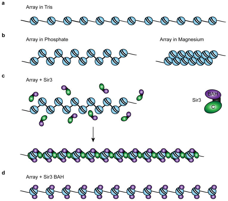
Figure 7. Model for a Sir3 chromatin fiber.
(a) Diagram of a 12-mer array in low-salt Tris buffer. (b) Arrays in 20 mM phosphate buffer pH 8.0 (containing ~40 mM Na+) are partially folded. Arrays in 1 mM MgCl2 buffer fold into 30 nm fibers. (c) Sir3 binds to arrays as a monomer, then subsequent dimerization via the Sir3 c-terminus bridges neighboring nucleosomes. Sir3 dimerization leads to array compaction distinct from 30 nm folding. (d) The Sir3 BAH domain binds to nucleosomes but cannot occlude linker DNA due to the absence of the C-terminal dimerization domain.

