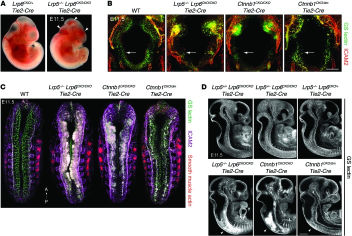
Figure 1. Effects of eliminating Ctnnb1 or Lrp5 and Lrp6 in ECs on vascularization of the E11.5 CNS.
(A) Bleeding in the hindbrain, midbrain, and forebrain of a Lrp5–/– Lrp6CKO/CKO Tie2-Cre embryo at E11.5 (arrowheads, right panel). Many embryos of this genotype also show bleeding in the spinal cord. Left, control embryo. (B) Transverse Z-stacked projections at E11.5 show severe vascularization defects in the Lrp5–/– Lrp6CKO/CKO Tie2-Cre and Ctnnb1CKO/CKO Tie2-Cre hindbrain and an intermediate vascularization defect in the Ctnnb1CKO/dm Tie2-Cre hindbrain (compare the regions highlighted by the arrows). Anti-ICAM2 stains ECs more strongly outside the CNS. GS lectin binds ECs and macrophages, which invade the hypovascularized CNS. Scale bar: 200 μm. (C) Spinal cord and hindbrain vascularization defects in E11.5 embryo whole mounts imaged from the dorsal surface. Z-stacks of approximately 70-μm thickness are shown. Smooth muscle actin highlights the somites. Lrp5–/– Lrp6CKO/CKO Tie2-Cre and Ctnnb1CKO/CKO Tie2-Cre embryos (center) show severe hypovascularization, formation of glomeruloid bodies, and bleeding within the spinal cord (white). The Ctnnb1CKO/dm Tie2-Cre embryo (far right) shows an intermediate vascularization defect. Anterior (A) is at the top; posterior (P) is at the bottom. (D) Optical sections in the sagittal plane of E11.5 embryos. The top 3 panels (Lrp5+/+ Lrp6CKO/CKO Tie2-Cre, Lrp5+/– Lrp6CKO/CKO Tie2-Cre, and Lrp5–/– Lrp6CKO/+ Tie2-Cre) show normal or nearly normal CNS vascularization. The left and middle bottom panels (Lrp5–/– Lrp6CKO/CKO Tie2-Cre and Ctnnb1CKO/CKO Tie2-Cre) show severe defects in CNS vascularization with bleeding that is most severe in the cervical spinal cord (arrowheads). The rightmost bottom panel (Ctnnb1CKO/dm Tie2-Cre) shows an intermediate vascularization defect in the spinal cord (arrowhead). Scale bar: 500 μm.

