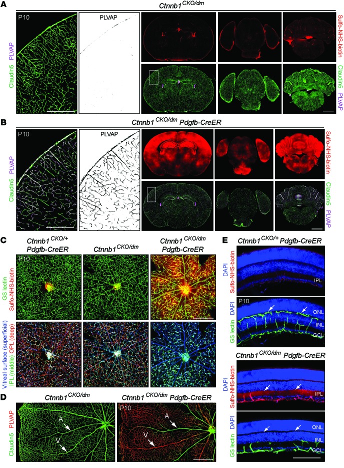
Figure 10. Transcriptionally inactive β-catenin cannot support normal retinal vascular development and BBB/BRB integrity.
(A and B) Brains from P10 Ctnnb1CKO/dm (control) and Ctnnb1CKO/dm Pdgfb-CreER mice treated with 60 μg 4HT at P6. Right panels, coronal sections at the anterior hippocampus (left), the pons (center), and the cerebellum (right). Enlarged images (left) show Claudin5 and PLVAP in the cerebral cortical vasculature, corresponding to the white rectangles (center). Ctnnb1CKO/dm brains show PLVAP–Claudin5+ vasculature and no sulfo-NHS-biotin leakage. Ctnnb1CKO/dm Pdgfb-CreER vasculature shows many PLVAP+Claudin5– ECs and extensive sulfo-NHS-biotin leakage. Scale bars: 500 μm (left panels); 2 mm (right panels). (C) Flat-mount retinas from P10 Ctnnb1CKO/+ Pdgfb-CreER, Ctnnb1CKO/dm, and Ctnnb1CKO/dm Pdgfb-CreER mice treated with 50 to 100 μg 4HT at P6. Upper row, the BRB is compromised in Ctnnb1CKO/dm Pdgfb-CreER retinas. Bottom row, retinal vasculature color coded by depth. Ctnnb1CKO/dm Pdgfb-CreER retinas have far fewer deep retinal capillaries. Scale bar: 400 μm. (D) Flat-mount retinas from P10 Ctnnb1CKO/dm (control) and Ctnnb1CKO/dm Pdgfb-CreER mice treated with 60 μg 4HT at P6. Ctnnb1CKO/dm Pdgfb-CreER retinas show efficient conversion of vein and capillary ECs from PLVAP–Claudin5+ to PLVAP+Claudin5–. Low levels of PLVAP in Ctnnb1CKO/dm capillaries are due to heterozygosity for Ctnnb1. Scale bar: 500 μm. (E) Cross sections of P10 retinas from control Ctnnb1CKO/+ Pdgfb-CreER (upper panels) and Ctnnb1CKO/dm Pdgfb-CreER mice (lower panels) treated with 50 to 100 μg 4HT at P6. Ctnnb1CKO/dm Pdgfb-CreER retinas show extensive vascular leakage in the IPL (arrows in the Sulfo-NHS-biotin panel) and lack deep retinal capillaries (arrows in the GS-lectin panels). Scale bar: 200 μm.

