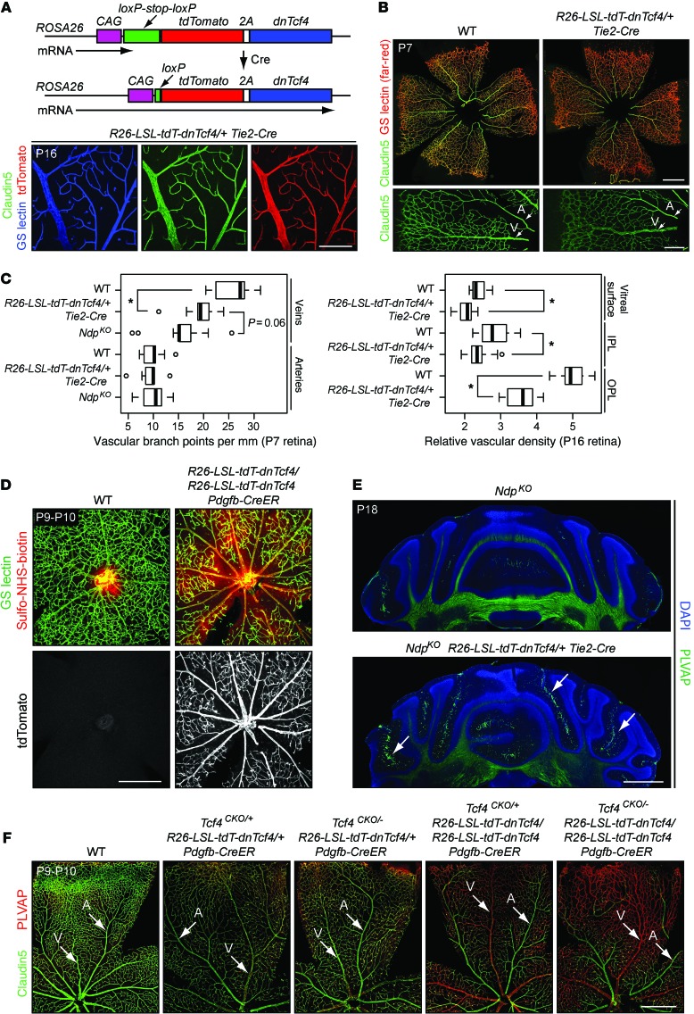
Figure 11. Production of dnTCF4 in ECs mimics the phenotypes seen with loss of canonical WNT signaling in the retina and cerebellum.
(A) The R26-LSL-tdT-dnTcf4 knockin before and after Cre-mediated recombination (upper panel). EC accumulation of tdTomato in R26-LSL-tdT-dnTcf4 Tie2-Cre retina flat mounts (lower panel). Scale bar: 200 μm. (B) P7 R26-LSL-tdT-dnTcf4/+ Tie2-Cre retinas show a modest decrease in Claudin5 expression in veins and capillaries compared with WT controls. Scale bar: 500 μm (upper panels); 200 μm (lower panels). (C) Quantification of branch points from retinal veins and arteries at P7 (left panel) and vascular density in the 3 retinal layers at P16 (right panel). *P < 0.01. (D) R26-LSL-tdT-dnTcf4/R26-LSL-tdT-dnTcf4 Pdgfb-CreER retinas (i.e., 2 alleles expressing dnTcf4) from P9–P10 mice treated with 50 μg 4HT at P3 show sulfo-NHS-biotin leakage and reduced vessel density (top panels). Pdgfb-CreER mediates nearly complete recombination at R26 as assessed by tdTomato fluorescence (bottom panels). Scale bar: 400 μm. (E) Genetic interaction between Ndp and R26-LSL-tdT-dnTcf4 with EC expression of dnTcf4. At P18, PLVAP+ ECs increase in the vasculature of the NdpKO;R26-LSL-tdT-dnTcf4/+ Tie2-Cre cerebellum (lower panel; white arrows) compared with the NdpKO cerebellum (upper panel). Scale bar: 1 mm. (F) Synergistic effect of reducing or eliminating Tcf4 and expressing different levels of dnTcf4 in ECs. Flat-mount P9–P10 retinas from mice that had received 40 μg 4HT at P1. With each reduction in Tcf4 or increase in dnTcf4, there are greater PLVAP expression and lower Claudin5 expression in veins and capillaries. Scale bar: 500 μm.

