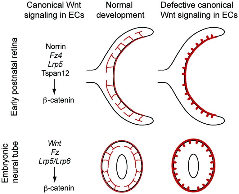
Figure 12. Schematic of angiogenesis defects in the neural tube and retina.
Top, early postnatal retina showing vascular invasion in WT versus a canonical WNT-signaling–deficient mutant (loss of Ndp or Fz4). Bottom, E11.5 neural tube showing vascular invasion in WT versus a canonical WNT-signaling–deficient mutant (loss of Ctnnb1 or Lrp5 and Lrp6). In the mutant cases, there is stunted invasion with the formation of glomeruloid bodies and hypertrophy of the surface vasculature. On the left, the currently defined components that mediate canonical WNT signaling are shown for retinal and neural tube angiogenesis.

