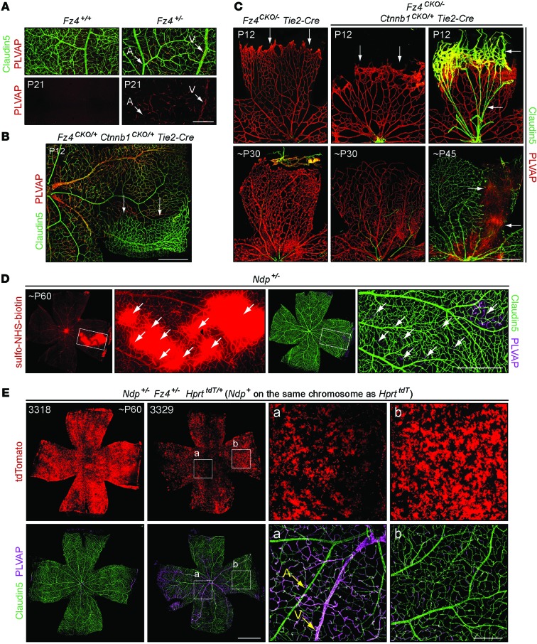
Figure 6. Vascular growth and BRB defects in retinas with different combinations of loss-of-function mutations in canonical WNT signaling components.
(A) PLVAP expression in capillary and vein ECs in Fz4+/– but not Fz4+/+ retinas. Scale bar: 200 μm. (B) Mosaic recombination in Fz4CKO/+ Ctnnb1CKO/+ Tie2-Cre retinas: recombined territory (upper two-thirds) with enhanced PLVAP and reduced Claudin5 in veins and capillaries and reduced vascular density, and unrecombined territory (arrows) with PLVAP–Claudin5+ ECs and normal vascular density. Scale bar: 500 μm. (C) More severe vascular defects in Fz4CKO/– Ctnnb1CKO/+ Tie2-Cre (right 4 panels) compared with Fz4CKO/– Tie2-Cre retinas (left 2 panels). Scale bar: 500 μm. (D) Representative flat-mount retina from an adult Ndp+/– female. Rare PLVAP+Claudin5– ECs (arrows) are associated with sulfo-NHS-biotin leakage. Boxed regions in low-magnification panels are enlarged to the right. Scale bar: 500 μm. (E) Compared with Ndp+/– retinas (e.g., D), approximately 50% of adult Fz4+/– Ndp+/– retinas show more extensive conversion of ECs from to a PLVAP+Claudin5– state. The HprttdT reporter and the Ndp+ allele are on the same X chromosome, and the Ndp– allele is on the unmarked X chromosome. tdTomato shows the pattern of X chromosome mosaicism. Retina 3318 shows few PLVAP+Claudin5– ECs. Retina 3329a shows multiple territories with a high density of PLVAP+Claudin5– ECs. Boxed regions marked a (low density of Ndp+ cells) and b (high density of Ndp+ cells) are enlarged at right. Scale bars: 1 mm (left panels); 200 μm (right panels).

