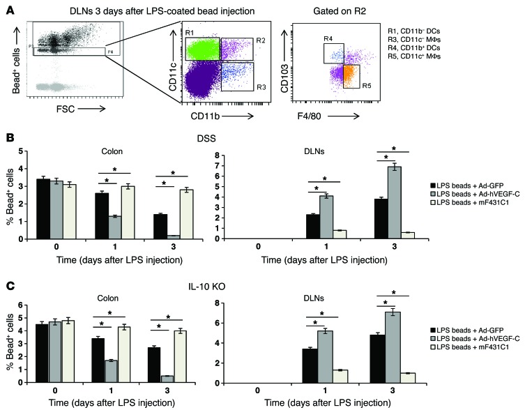
Figure 6. Systemic delivery of Ad-hVEGF-C accelerates bacterial antigen clearance from the inflamed colon to DLNs.
(A–C) A single intramucosal injection of carboxylated crimson fluorescent LPS-coated beads was administered on day 5 after the second DSS cycle (chronic inflammation) and on day 21 after the first Ad-hVEGF-C/mF431C1 administration to IL-10–KO mice. Colitic mice treated with Ad-GFP and injected with LPS-coated beads were used as controls. (A) FACS plots showing DCs (R1 and R4) and MΦs (R3 and R5) within the bead+ cell population based on the expression of CD11b (R3), CD11c (R1), F4/80 (R5, gated on R2), and CD103 (R4, gated on R2). (B and C) Clearance of single-bead+ cells from the inflamed colon (left panels) to the DLNs (right panels) was monitored, and quantification is reported as the mean per group ± SEM. n = 5 mice per group. *P < 0.05 versus LPS-coated beads plus Ad-GFP.

