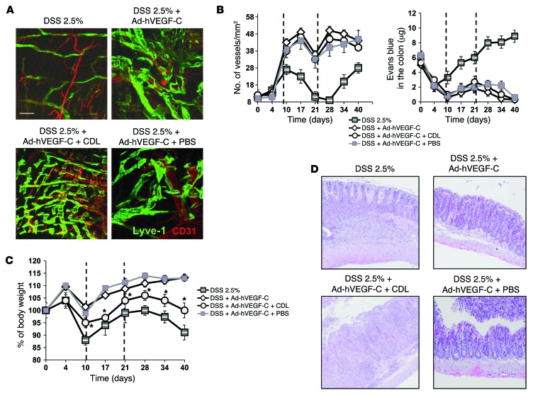
Figure 7. MΦ depletion using CDL reduces protection of VEGF-C–overexpressing mice during DSS-induced colitis.
Mice undergoing 2 cycles of DSS treatment were injected with Ad-hVEGF-C and intrarectally administered CDL or PBS, as described in Methods. (A) Representative images of whole-mount staining of distal colons from the indicated groups after the second cycle of DSS (day 28). Scale bar: 75 μm. (B, left panel) LYVE1+ vessels were counted in 10 regions per section, and the numbers were normalized per total section area expressed in mm2. Results are presented as the mean vessel density per group ± SEM. (B, right panel) Evans blue was extracted 16 hours after the dye injection from distal colons of comparable weight. Graph shows the total dye remaining in the colon expressed in μg. Data represent the mean per group ± SEM (n = 4–6/group). (C and D) CDL administration significantly reduced the protective effect of VEGF-C on the basis of the percentage of body weight (C) and histological examination (D) compared with control mice, which received only DSS plus VEGF-C. Black dashed lines represent the 2 DSS cycles. (D) Representative H&E-stained images of colons from the indicated groups of mice at the end of the experiment (day 40). Original magnification, ×20. Results in C are presented as the mean value per group at the indicated time points ± SEM (n = 4–6/group). *P < 0.05 versus DSS plus VEGF-C alone.

