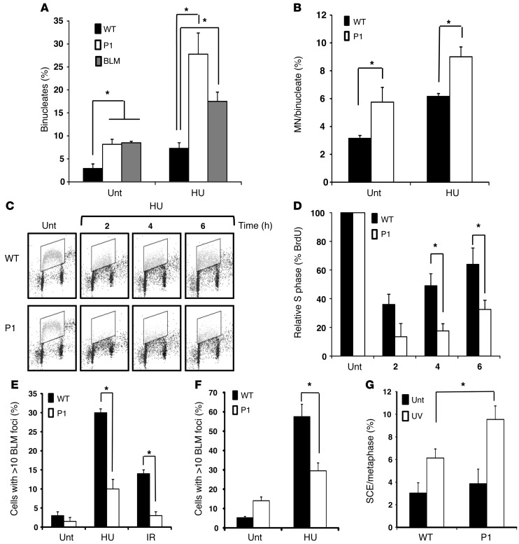
Figure 4. Increased micronucleus formation, impaired S phase progression, and increased SCE in LCLs with NSMCE2/MMS21 mutations.
(A) Binucleates in LCLs from WT, P1, and from a Bloom syndrome patient, either untreated (Unt) or following repeated exposure to HU (50 μM/d for 4 days). (B) HU-induced MN formation in Cyt-B–induced binucleate LCLs. (C) BrdU flow cytometry profiles from WT and P1 LCLs either untreated or following treatment with low-dose HU (250 μM). S phase content (boxed area) following a BrdU pulse 30 minutes prior to each time point is shown. (D) Normalized total S phase content compared with that of untreated cells at each time point. Means ± SD are shown (n = 3). (E) BLM foci formation in P1 LCLs relative to WT following treatment with HU (1 mM for 16 hours) or IR (10 Gy for 16 hours). (F) BLM foci formation in primary dermal fibroblasts from P1 compared with WT following HU exposure (1 mM for 24 hours). (G) UV radiation–induced SCEs in WT and P1 LCLs compared with untreated cells. These effects, although significant, were not as profound as those observed in BLM patient LCLs, which exhibited 23 ± 6 SCEs/metaphase in untreated cells and 38 ± 4 SCEs/metaphase following UV irradiation. *P < 0.05 (unpaired t test).

