Figure 1.
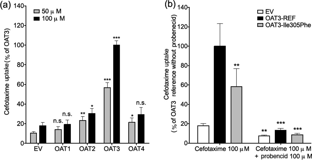
Uptake of cefotaxime in HEK293 Flp-In cells expressing organic anion transporters (OATs). (a) Uptake of cefotaxime (50 µM and 100 µM) in HEK293 Flp-In cells expressing human OAT1, OAT2, OAT3 and OAT4. The cells were exposed to cefotaxime for 5 minutes. Significantly greater cefotaxime (50 µM and 100 µM) uptake was observed in HEK293-Flp-In cells transfected with OAT3 compared to cells transfected with empty vector (EV), OAT1, OAT2 and OAT4 (p< 0.005). Asterisks indicate significantly different uptake values from EV controls. Note: * p-value < 0.05, ** p-value < 0.01, *** p-value < 0.005, n.s. not-significant. Multiple comparisons were analyzed using one-way analysis of variance followed by Dunnett’s two-tailed test. Data are shown as mean ± SD from 2 repeated experiments, and each experiment was performed in triplicate. Results are expressed as the percent of activity of the OAT3-reference. (b) Uptake of cefotaxime (100 µM) with and without the OAT inhibitor, probenecid (100 µM), in HEK293 Flp-In cells expressing OAT3 reference (OAT3-REF) and OAT3 variant, Ile305Phe. The cells were exposed to cefotaxime with or without probenecid for 5 minutes. The result showed significantly reduced cefotaxime uptake in cells transfected with OAT3 Ile305Phe compared to OAT3 reference, ** p-value < 0.01. In the presence of probenecid, cefotaxime uptake in cells transfected with empty vector (EV), OAT3 reference (OAT3-REF) or OAT3 variant (OAT3-Ile305Phe), were significantly reduced compared to cells without probenecid, ** p-value < 0.01, *** p-value < 0.005. Data are shown as mean ± SD from 2 repeated experiments and each experiment was performed in triplicate. Results are expressed as the percent of activity of the OAT3-reference (OAT3-REF).

