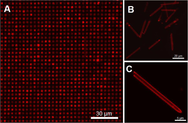
Figure 4.

Fluorescence confocal images of PEM-coated and DOX-loaded micropillars. Fluorescence confocal micrograph of the micropillar arrays in top view after PEM coating (eight bilayers) and DOX loading (A); detached hollow micropillars with uniform size distribution (B); and single detached micropillar with PEM and DOX all along the walls (C).
