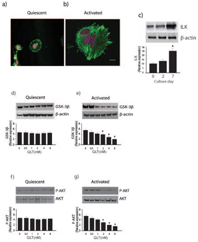
Figure 1. Differential ILK activity in quiescent and activated stellate cells.

In (a) and (b), stellate cells were isolated from normal rats livers as in Methods, allowed to adhere to glass-coated culture dishes, and grown in 20% serum-containing medium for 1 day (a, quiescent) or 7 days (b, activated). Cells were fixed, and immunohistochemistry was performed as described in Methods. Representative images are shown (ILK is labeled with AL-488 (green)), and nuclei are labeled with DRAQ5 (pink)). The bar shown in the lower right hand corner of (a) represents 10 microns. In (c), stellate cells were as in (a/b); cell lysates were subjected to immunoblotting (50 μg total protein) with anti-ILK antibody. Representative blots are shown in the top portion of the panel and in the bottom portion of the panel, specific bands were scanned and quantitated, and presented graphically (n = 4; *p < 0.05, vs. day 0 or day 2). In (d), quiescent cells were exposed to serum overnight and were exposed to the indicated concentrations of QLT-0267 in medium containing 0.1% serum for 18 hours. Lysates were harvested and GSK3-β phosphorylation was detected by immunoblotting as in Methods. Signals were scanned, normalized to the control value, and displayed in the graph below the immunoblot (n=3). In (e), activated cells and sample processing were as in (d), (n=3, *p < 0.05, compared to control - without QLT). In (f/g), cells were as in (d/e) (i.e. quiescent and activated cells, respectively), and phospho-Akt was detected by immunoblotting as in Methods (n=3, *p < 0.05, compared to control - without QLT).
