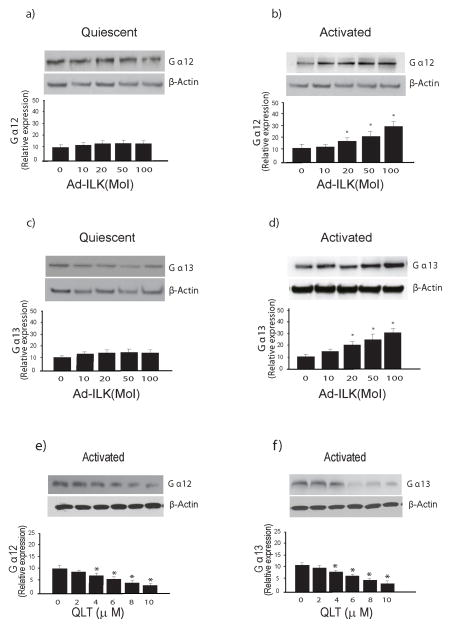
Figure 7. ILK modulates expression of Ga12 and Ga13 in activated stellate cells.
In (a) through (d), stellate cells were as in Figure 1; each quiescent (cultured for one day) and activated cells (cultured for 7 days) were exposed to adenovirus-expressing ILK (Ad-ILK) for 24 hours. Forty-eight hours later, cell lysates (50 μg total proteins) were subjected to immunoblotting to detect Gα12 (a/b) or Gα13 (c/d). Representative immunoblots are shown in the upper panels, and below them, a stripped blot re-probed for β-actin; subsequently, specific bands were quantified, normalized to the signal for β-actin and presented graphically (n = 3; *p<0.05, compared to “0”). In (e) and (f), activated stellate cells were exposed to different concentrations of QLT for 18 hours in medium containing 0.1% serum, cell lysates were harvested (50 μg total protein) and subjected to immunoblotting to detect Gα12 and Gα13. A representative immunoblot is shown in the upper panel, and below it, a stripped blot re-probed for β-actin; specific bands were quantified, normalized to the signal for β-actin and presented graphically (n = 3; *p<0.05, compared to “0”).

