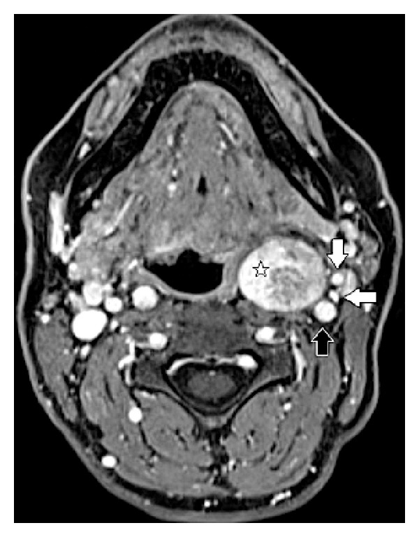
Figure 2.

Patient 2, T1-weighted enhanced MRI scan: ovoid mass occupying the left retrostyloid compartment, 3 × 4 cm in size (white star). The internal carotid artery (black arrow), the internal jugular vein (horizontal white arrow), and the external carotid artery (vertical white arrow) are all displaced laterally.
