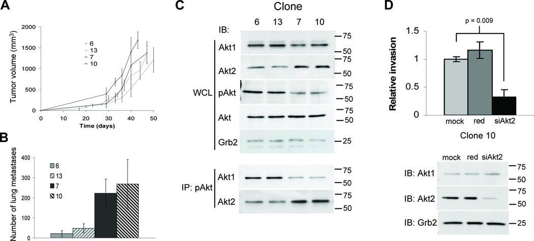
Figure 3.
Elevated Akt2 levels increase invasion and metastasis. A, Cells were injected into the mammary fat pad of athymic mice and primary tumors measured twice weekly. Tumor volumes represent the mean for 5 independent mice (+/− SD). B, Hematoxylin and eosin stained lungs were scored for the number of metastases. The values represent the mean number for 5 individual mice (+/− SD). C, Immunoblot analyses of Akt1, Akt2, pAkt and total Akt of TM15 clone whole cell lysates (WCL). To examine Akt isoform activation, pAkt (Ser473) was immunoprecipitated from lysates and immunoblots performed for Akt1 and Akt2. D, Invasion through Matrigel was assayed for mock, red negative control and Akt2-specific siRNA transfected TM15 clone 10. Pixel count analyses of Crystal Violet stained membranes were performed and relative invasion determined with mock transfection (mock) control set to 1. Values represent the mean of assays performed in triplicate (+/− SD) and statistical analyses performed using a student’s t-test. Immunoblots for Akt1 and Akt2 were performed on cell lysates, with Grb2 detected as a control for loading.

