Figure 5.
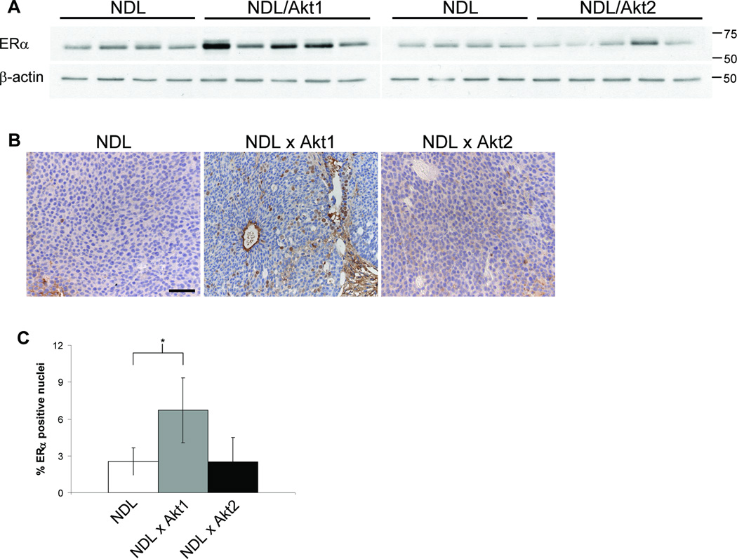
NDL/Akt1 mammary tumors express elevated ERα. A, Lysates of NDL, NDL/Akt1 and NDL/Akt2 mammary tumors were immunoblotted for ERα. β-actin was detected as a control for loading. The NDL lysates were identical for the left and right panels allowing direct comparison. B, Representative images of tumor sections stained with ERα-specific antibody. Bar, 50µm. C, The percentage of ERα positive nuclei was determined for 10 fields for each tumor section and represents the mean of 4 independent tumors for each genotype (+/− SD). *, P = 0.045 (student t-test).

