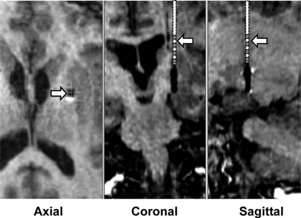
FIG. 1.
Postoperative MRI scans showing the projected location of the DBS lead during putamen DBS. The reconstruction was based on an MRI scan, with the DBS lead in its final location in the GPi. MRI shows the location of the DBS contacts in the GPi. The putamen DBS was in the same trajectory with the distal edge of the distal contact 10 to 13 mm above the location of the distal edge of the distal contact with the DBS lead in the GPi. Estimated positions of the DBS lead, shown schematically, in the coronal and sagittal plane are indicated by the arrows. Location of the putamen DBS lead in the axial section is indicated by the arrow. As can be seen, during putamen DBS, the lead was in the posterior medial region of the putamen.

