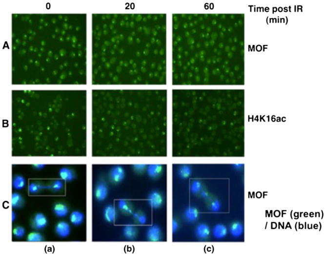
Fig. 7.
Male X-chromosome staining for A MOF or B H4K16ac at different time points post-irradiation in SL-2 cells. C Cells showing the status of male X chromosomes detected by MOF staining (MOF color is green and DNA is blue): a daughter cells showing similar intensity of MOF staining of X chromosomes at late anaphase; b one daughter cell showing MOF staining whereas the other lacks MOF staining at telophase; and c two daughter cells sharing three MOF signals at telophase

