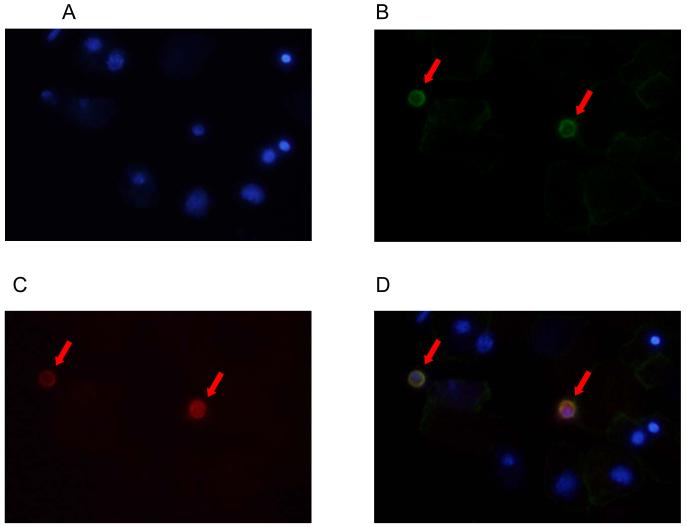
Figure 3. Double-label immunofluorescence staining of an amniotic fluid cell pellet from a patient with intra-amniotic infection/inflammation using antibodies to endoglin and CD14.
(A) DAPI (4′,6-Diamidino-2-Phenylindole, Dihydrochloride) staining of nuclei (blue). (B) CD14 expression on macrophages (green; Alexa Fluor 488). (C) Endoglin expression on macrophages (red; Alexa Fluor 594). (D) The merged image showing that macrophages expressed both CD14 (green) and endoglin (red).

