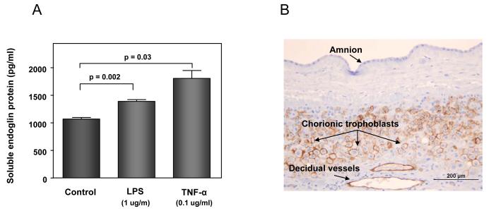
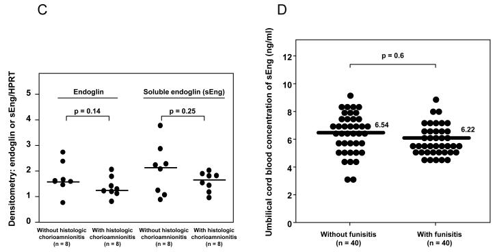
Figure 7. Potential sources of an increased soluble endoglin (sEng) in the amniotic fluid of patients with intra-amniotic infection/inflammation.
(A) amniotic fluid macrophages: the mean concentration of sEng in supernatant of U937-derived macrophages was significantly higher than in the control after treatment with LPS (p=0.002) or TNF-α (p=0.03). Data represents mean ± SEM. All the experiments were done in triplicate. The p-values were calculated by Student’s t-tests. (B) chorioamniotic membranes: immunohistochemical staining of chorioamniotic membranes demonstrated that endoglin was expressed in chorionic trophoblasts and endothelial cells of decidual vessels, but not in amnion cells. (C) chorioamniotic membranes: densitometric analysis of chorioamniotic membranes for the expression of endoglin and sEng over HPRT (hypoxanthine-guanine phosphoribosyltransferase) between patients with preterm labor (PTL) with (n=8) and without (n=8) histologic chorioamnionitis. There was no significant difference in the median endoglin density ratio and sEng density ratio between patients with and without histologic chorioamnionitis [endoglin density ratio: median 1.24, interquartile range (IQR) 1.14–1.58 vs. median 1.58, IQR 1.49-2.0, p=0.14; sEng density ratio: median 1.66, IQR 1.29–1.83 vs. median 2.13, IQR 1.14–2.55, p=0.25]. (D) umbilical cord blood: there was no significant difference in the median plasma concentration of sEng in the umbilical cord blood of neonates born to mothers with PTL with (n=40) and without (n=40) histologic chorioamnionitis/funisitis [median 6.22 ng/mL, IQR 5.65-7.33 ng/mL vs. median 6.54 ng/mL, IQR 5.62-7.51 ng/mL; p=0.6].


