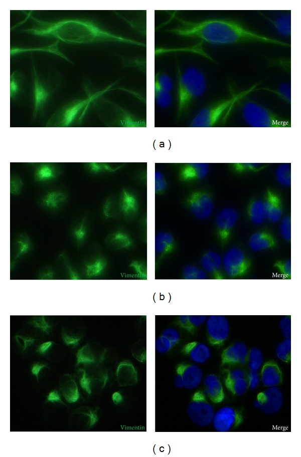
Figure 3.

Immunofluorescence images of vimentin in MDA-MB-231. Immunostaining of vimentin (green color) and HOECHST 33342 to stain nuclei (blue color) after 24 hours in on ground control cells (a), RPM adherent cells (b), and RPM cell clumps (c). Magnification ×400.
