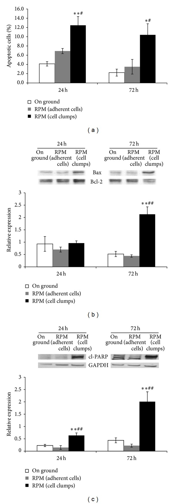
Figure 5.

Apoptosis analysis in MDA-MB-231. Apoptotic rate in RPM cultured MDA-MB-231 and on ground cells was determined by a dual parameter flow cytometric assay (a). Histograms show the percentage of apoptotic cells (Annexin V+/7-AAD-); each column represents the mean value ± SD of three independent experiments. Immunoblot bar chart showing the expression of Bax/Bcl-2 ratio (b) and cleaved PARP (c) in on ground control cells, RPM adherent cells, and RPM cell clumps at 24 and 72 hours. Columns and bars represent densitometric quantification of optical density (OD) of specific protein signal normalized with the OD values of the GAPDH served as loading control. Each column represents the mean value ± SD of three independent experiments. *P < 0,05; **P < 0,01 versus on ground control; # P < 0,05; ## P < 0,01 versus RPM adherent cells by ANOVA followed by Bonferroni post-test.
