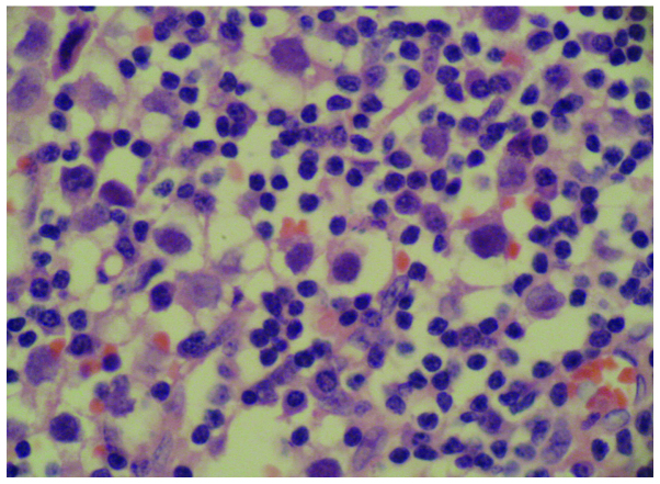
Figure 2.
Dysgerminoma (magnification, ×400, hematoxylin and eosin staining). The tumor cells are large in size and round or ovoid in shape, and have distinct borders. The nucleus at the center of the cell is large and round, and nuclear division is often observed. There is abundant transparent cytoplasm. Lymphocyte infiltration is observed in the connective tissue.

