Fig. 1.
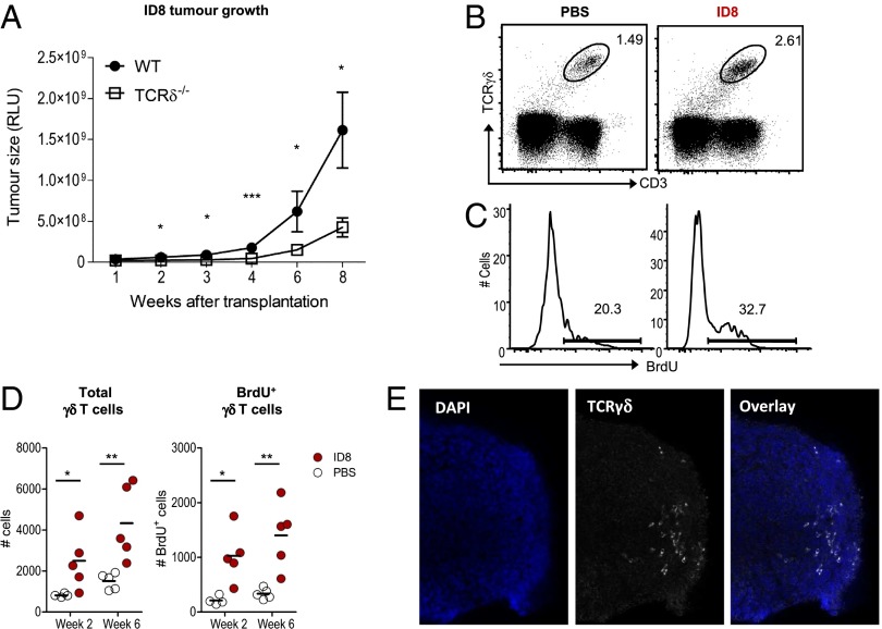
γδ T cells infiltrate ID8 tumors and enhance ovarian cancer cell growth in vivo. (A) ID8 tumor growth in C57BL/6 WT (n = 6) and TCRδ−/− female mice (n = 8), measured by luciferase bioluminescence at the indicated weeks posttransplantation. (B) Representative FACS plots for γδ T cells in peritoneal exudates of ID8-bearing mice or PBS controls (at week 6 postinoculation). (C and D) Absolute numbers of total and BrdU+ γδ T cells in ID8-bearing mice or PBS controls. BrdU was provided during a period of 2 wk before analysis. Each dot represents one animal. (E) Representative immunofluorescence imaging of γδ T cells in ID8 tumor foci. Data are representative of three independent experiments; *P < 0.05, **P < 0.01, ***P < 0.001.

