Fig. 3.
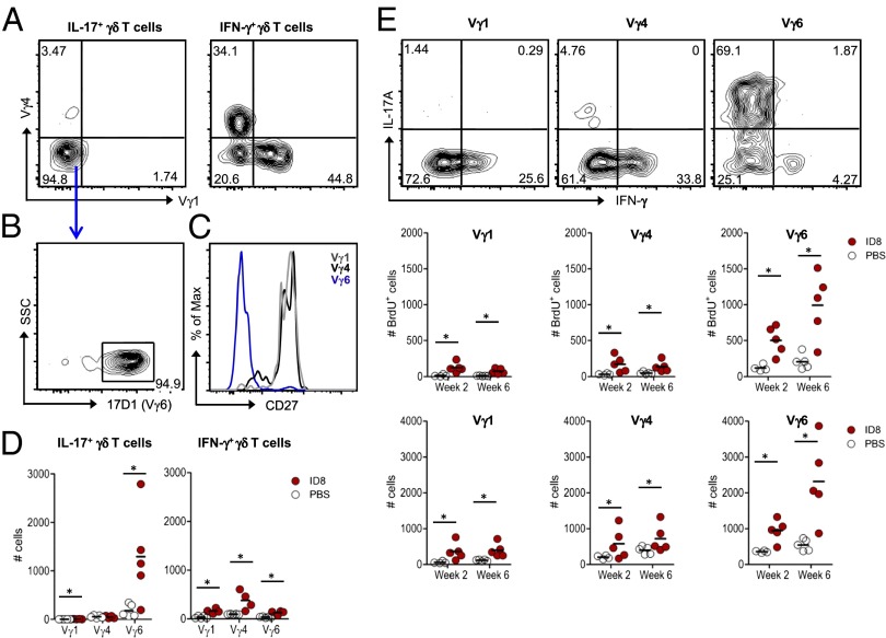
IL-17 production by γδ T cells is essentially restricted to a distinctive CD27(−) Vγ6(+) cell subset. Peritoneal exudates were analyzed at week 2 and week 6 postinoculation of ID8 cells or PBS. (A) Representative FACS plots of Vγ1 and Vγ4 stainings within IL-17A(+) or IFN-γ(+) γδ T cells in tumor-bearing mice (at week 6). (B) FACS staining with GL3 and 17D1 monoclonal antibodies to detect Vγ6(+) γδ T cells. (C) Histogram overlay of CD27 staining in Vγ1, Vγ4, and Vγ6 subsets of γδ T cells in tumor-bearing mice (at week 6). (D) Absolute numbers of Vγ1, Vγ4, and Vγ6 subsets of IL-17A(+) or IFN-γ(+) γδ T cells in ID8 tumor-bearing mice or in PBS-injected controls (at week 6). Each dot represents one animal. (E) Representative intracellular IL-17A and IFN-γ stainings (Top), BrdU incorporation (Middle), and absolute numbers (Bottom) of Vγ1, Vγ4, and Vγ6 subsets of γδ T cells in ID8 tumor-bearing mice or in PBS-injected controls (at weeks 2 and 6). Data are representative of three independent experiments; *P < 0.05.

