Fig. 2.
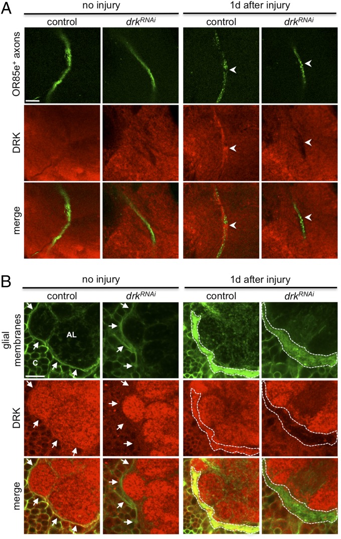
DRK is recruited to glial membranes surrounding the injured axons. (A) Endogenous DRK (red, α-DRK) in glia was recruited to the severed mp nerves labeled with OR85e-mCD8::GFP (green, α-GFP) 1 d after axotomy. Before injury, DRK expression was evenly distributed in the SOG but not colocalized with the OR85e+ maxillary nerve. One day after mp injury, DRK expression was increased around the degenerating maxillary nerves (arrowheads), which was not seen in drkRNAi animals. Representative images (single slice) are shown. (Scale bar: 10 μm.) (B) Endogenous DRK expression in glia was increased 1 d after axotomy. Glial membranes were labeled with mCD8::GFP by glia-specific repo-Gal4 driver. Antennal ablation (removal of the third segment of antennae) was performed to induce a greater extent of axon degeneration in the antennal lobe (AL). Normally, thin glial membranes ensheath (arrows) the antennal lobe and each glomerulus (no injury, control). However, 1 d after antennal ablation, ensheathing glia became hypertrophy (1 d after injury, dashed lines), and strong DRK immunoreactivity (red) was found in hypertrophic region of ensheathing glia (yellow), but not when drkRNAi was expressed by repo-Gal4, indicating that the increase of DRK is glia-specific. Representative images (single slice) are shown. C, cortex. (Scale bar: 10 μm.)

