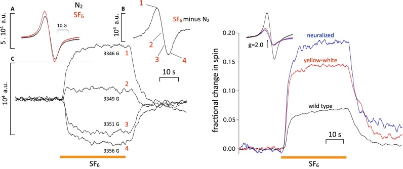
Fig. 2.
(Left, Inset A) Spin signal from ∼30 W1118 WT flies before (black trace) and after (red trace) exposure to SF6. (Left, Inset B) Difference trace resulting from the two traces in Left, Inset A (i.e., the spin signal introduced by SF6). (Left, Inset C) Four traces measured at fixed values of magnetic field corresponding to points 1–4 on trace B (i.e., 1, near the top; 2, close to the cross-over; 3, halfway down; 4, just past the bottom peak). The amplitude of the traces in Left, Inset C matches the amplitudes expected from Left, Inset B, showing that the spin changes seen in fixed field continuous measurements do, indeed, reflect an underlying resonance. The other anesthetics behave similarly. (Right) Effect of melanin content in different fly strains on anesthetic-induced spin changes. (Inset) Intrinsic spin signals of W1118 WT and two pale fly strains [yellow-white (red trace) and neuralized (blue trace)] in which eumelanin is absent. The intrinsic spin signal of the pale strains is much smaller, which was expected. Percentage change in spin in response to SF6 in the three strains. If the signal was caused by melanin, it should either be reduced in amplitude or disappear altogether. Instead, it increases in inverse proportion to the intrinsic spin level, showing that the two are independent.

