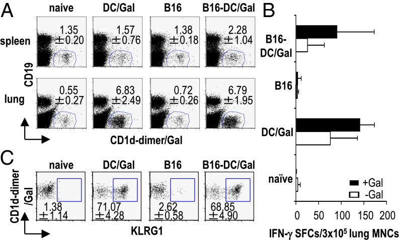
Fig. 1.
Prolonged activation of iNKT cells in the lung by an injection of DC/Gal. B16 melanoma cells (2 × 10⁵ cells per mouse) were administered i.v. to C57BL/6 mice. On day 7, B16-bearing or naive mice were immunized with 1 × 10⁶ DC/Gal. (A) The frequency of iNKT cells in spleen and lung was assessed in the four groups of mice on day 14 (n = 4; data are shown as mean ± SEM). (B) The lung MNCs were isolated on day 14 from the four groups of mice and cultured for 16 h in the presence or absence of α-GalCer (100 ng/mL). IFN-γ production by lung MNCs was measured by ELISPOT assay (n = 4–6; data are shown as mean ± SEM). (C) KLRG1 expression on lung iNKT cells was analyzed by gating on CD19−CD1d-dimer/Gal+ cells (n = 5; data are shown as mean ± SEM).

