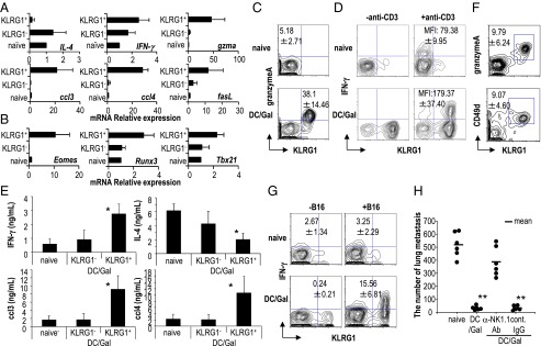
Fig. 3.

Characterization of KLRG1+ iNKT cells in DC/Gal-injected mice. C57BL/6 mice were immunized with DC/Gal. One month later, KLRG1+ and KLRG1− iNKT cells in the lung were purified by FACS Aria. As a control, iNKT cells in the lung of naive mice were shown as naive iNKT cells. (A and B) Quantitative analyses of gene expression were performed by quantitative real-time PCR using the primer sets shown in Table S1 (n = 4; data are shown as mean ± SEM). (C and D) To examine the functional features of KLRG1+ iNKT cells, the expression of granzyme A (C) and the production IFN-γ (D) in iNKT cells in the lung were analyzed. For intracellular staining of IFN-γ, lung MNCs were stimulated with anti-CD3 Ab plus anti-CD28 Ab for 2 h in the presence of brefeldin A. Analysis gates were set on CD19−CD1d-dimer/Gal+ cells (n = 4–6; data are shown as mean ± SEM). (E) The supernatants from the cultures with anti-CD3 Ab plus anti-CD28 Ab for 24 h were analyzed for IFN-γ, IL-4, CCL3, and CCL4 (n = 4; data are shown as mean ± SEM). *P < 0.05 naive iNKT or KLRG1− iNKT cells versus KLRG1+ iNKT cells. (F) The expression of granzyme A and CD49d of KLRG1+ iNKT cells in the lung was analyzed 6 mo after immunization with DC/Gal (n = 4; data are shown as mean ± SEM). (G and H) Mice that had been immunized with DC/Gal were challenged with B16 melanoma cells 4 mo later. In some experiments, the mice were treated with control rat IgG or anti-NK1.1 Ab. IFN-γ secretion assay for B16 reactive iNKT cells was performed 12 h later in mice vaccinated with or without DC/Gal (G). Antitumor effects were evaluated 2 wk later by counting the number of metastases in the lungs (H) (n = 4–6; data are shown as mean ± SEM). **P < 0.01 naive versus DC/Gal, anti-NK1.1 Ab treatment (DC/Gal) versus control rat IgG treatment (DC/Gal).
