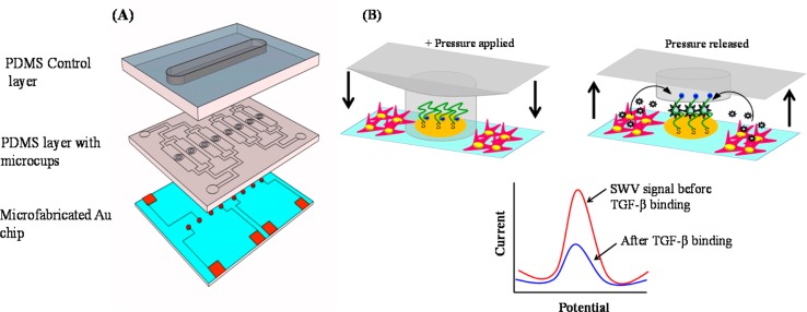
Scheme 1. (A) Microfluidic Device Was Composed of Three Layers: Glass Slide with Micropatterned Au Electrodes, PDMS Layer with Fluid Channels and Microcups, and Another PDMS Layer for Controlling of Microcups. (B) Diagram Showing Actuation of Microcups to Protect Electrodes during Collagen Coating and Cell Seeding into the Channel.

Once cells are activated and microcups are raised, the electrodes may be used for detection of TGF-β1. Redox labeled aptmer molecules immobilized on the electrode interact with TGF-β1, leading to a change in redox current. The redox current is decreased when TGF-β1 binds.
