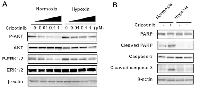
Figure 2.

Western blot analyses of a downstream signal and apoptosis-related molecules in the H3122 cell line. (A) Downstream signal. The cells were incubated under normoxia or hypoxia for 48 h, then crizotinib was added three hours before the sample collection. The concentrations of crizotinib were 0, 0.01, 0.1 and 1 μM. The phosphorylation of AKT and ERK was dose-dependently reduced by crizotinib under normoxia. Under hypoxia, however, the phosphorylation was less reduced by crizotinib. β-actin was used as an internal control. (B) Apoptosis-related molecules. The samples were collected 48 h after DMSO (control) or crizotinib stimulation under normoxia or hypoxia. The concentration of crizotinib was 0.1 μM. The expression of both cleaved PARP and cleaved caspase-3 was elevated by crizotinib under normoxia, whereas neither expression level was elevated under hypoxia. β-actin was used as an internal control.
