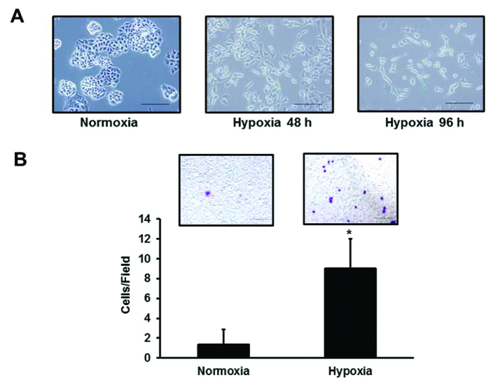
Figure 3.
Morphologic change and migration of the H3122 cell line. (A) Morphologic change. Hypoxia time-dependently induced morphologic changes that are characteristic of the EMT, including cell scattering and an elongation of the cell shape. Scale bar, 20 μm. (B) Migration. The migration assays were performed using the Boyden chamber method. After incubation for 48 h under normoxia or hypoxia, the cells that had migrated to the outer side of the membranes were fixed and stained with crystal violet staining solution, then counted using a light microscope. The experiment was performed in triplicate. Under hypoxia, the number of migrating cells was significantly higher than that under normoxia (8.67±3.5 vs. 1.33±1.53/Field, *P=0.026). Columns, mean of independent triplicate experiments; error bars, SD; *P<0.05.

