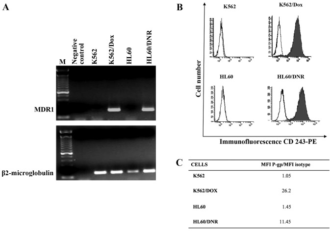
Figure 2.
Expression of P-gp by the leukemic cells. MDR1 mRNA expression was studied in K562, K562/Dox, HL60 and HL60/Dnr cells by RT-PCR using β2-microglobulin as control (A). Flow cytometry analysis of P-gp in these cells using anti-CD243-phycoerythrin (B). P-gp expression was quantified as the mean fluorescence intensity (MFI) shift (ratio of the MFI of P-gp and isotype control) (C).

