Figure 5.
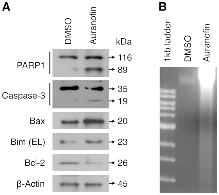
Auranofin induces cellular apoptosis in SKOV3 cells. (A) SKOV3 cells were treated with auranofin (100 nM) or control (DMSO) for 48 h. Total lysates of cells were analyzed by western blot analysis with specific antibodies against apoptosis-related proteins as indicated. β-actin represents the loading controls. (B) DNA samples extracted from SKOV3 cells, which were treated with auranofin or control as described above, and subjected to DNA fragmentation assay. Equal amounts of the extracted DNA (2 μg/lane) and size markers (1-kb ladder) were subjected to electrophoresis on 2% agarose gels, which were stained with ethidium bromide and photographed.

