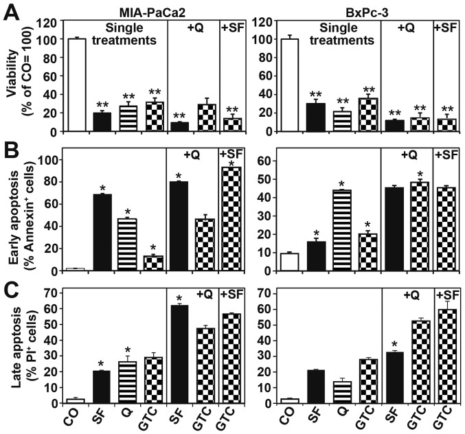
Figure 4.
Combinations of dietary agents are superior in reducing viability and inducing apoptosis compared to single agents. (A) MIA-PaCa2 and BxPc-3 cells were treated as described in Fig. 2B. Ninty-six hours later viability was detected by the MTT assay. (B) Apoptosis was measured by annexin staining, followed by FACS analysis. The percentage of annexin-positive cells is shown. (C) Apoptosis was measured by staining of the cells with propidium iodide followed by FACS-analysis. The data were quantified and statistically analyzed as described in Fig. 2.

